Reviewed for accuracy against published gout and heel-pain guidance available through October 2026.
A painful heel can make the first step out of bed feel impossible. Sometimes the problem comes from a tendon or the thick band of tissue under the foot. Other times, a sudden attack of swelling and tenderness raises a different question: could gout be responsible? Although gout is famous for affecting the big toe, urate crystals can also accumulate in joints and soft tissues around the heel. The location of pain alone cannot tell you which condition is present.
Gout in the heel is possible, particularly near the back of the heel, ankle, bursae, or Achilles tendon. It is less common than a classic big-toe flare. A new hot, swollen, very painful area deserves assessment, especially if fever or illness is present, because infection can resemble gout and requires different treatment.
Can You Get Gout in Your Heel?
Yes. Gout develops when monosodium urate crystals trigger inflammation. The heel is a region rather than a single joint: it contains the calcaneus (heel bone), the Achilles tendon insertion, bursae, soft tissues and neighboring joints. Crystal deposits may occur in some of these structures, including in people with longstanding or tophaceous gout. That does not mean every sore heel is gout.
Some patients ask, “Can you get gout in the heel of your foot even if your big toe feels normal?” You can. A flare need not begin in the big toe, and different attacks can involve different locations. Clinicians consider the timing, appearance, previous attacks and diagnostic findings rather than assuming all heel symptoms have the same cause.
What Does Gout in the Heel Feel Like?
A typical acute gout flare develops quickly, often overnight. Pain can escalate over hours and make contact from a sock or bedsheet uncomfortable. The involved area may be swollen, warm and visibly red, although redness can be less obvious on darker skin. Patients may find it hard to walk or wear shoes. Some attacks resolve over several days; others persist longer without effective treatment.
A deep ache that has slowly increased over weeks, particularly with running or repeated loading, may instead suggest a mechanical heel problem. Gout and mechanical problems can coexist, so pain quality is only one clue. People with chronic tophi may notice firm nodules with less dramatic inflammation between flares.
Sudden pain, warmth, and sensitivity
An abrupt onset of pronounced tenderness is consistent with an inflammatory process. It cannot distinguish gout from bacterial infection, however. Fever, feeling unwell, or rapidly spreading changes deserve urgent medical evaluation.
Swelling and trouble bearing weight
Swelling around the ankle or back of the heel can make the foot stiff. A patient may limp or avoid placing the heel against a shoe. Inability to bear weight, especially after injury or accompanied by significant redness, should not be self-diagnosed.
Gout Heel Pain Location: Back, Bottom, and Sides of the Foot
Gout at the back of the heel
The Achilles tendon attaches to the rear of the calcaneus. Gouty tophi can develop in or near tendons and adjacent soft tissue, sometimes producing a firm bump, pain with shoe pressure, or swelling. More commonly, pain here comes from insertional Achilles tendinopathy, retrocalcaneal bursitis or friction from footwear. An abrupt pop, weakness or inability to push off the foot raises concern for tendon injury and calls for prompt care.
Gout under the heel
The underside of the heel is a frequent site of plantar fasciitis. People often describe sharp first-step pain after getting out of bed or rising from prolonged sitting. Gout can affect nearby structures, but an isolated ache precisely under the heel is not a reliable sign of gout. A careful examination can determine which tissues are tender and whether inflammatory findings are present.
Around the ankle or sides of the heel
A flare in the ankle or adjacent joints may be experienced as heel pain because swelling spreads or because the location is difficult to pinpoint. Pain can also arise from bursitis, tendon conditions, nerve irritation or injury. Medical assessment becomes more important when symptoms are new, severe or atypical for a person’s previous gout attacks.
Pictures of Gout in the Heel: What Can They Show?
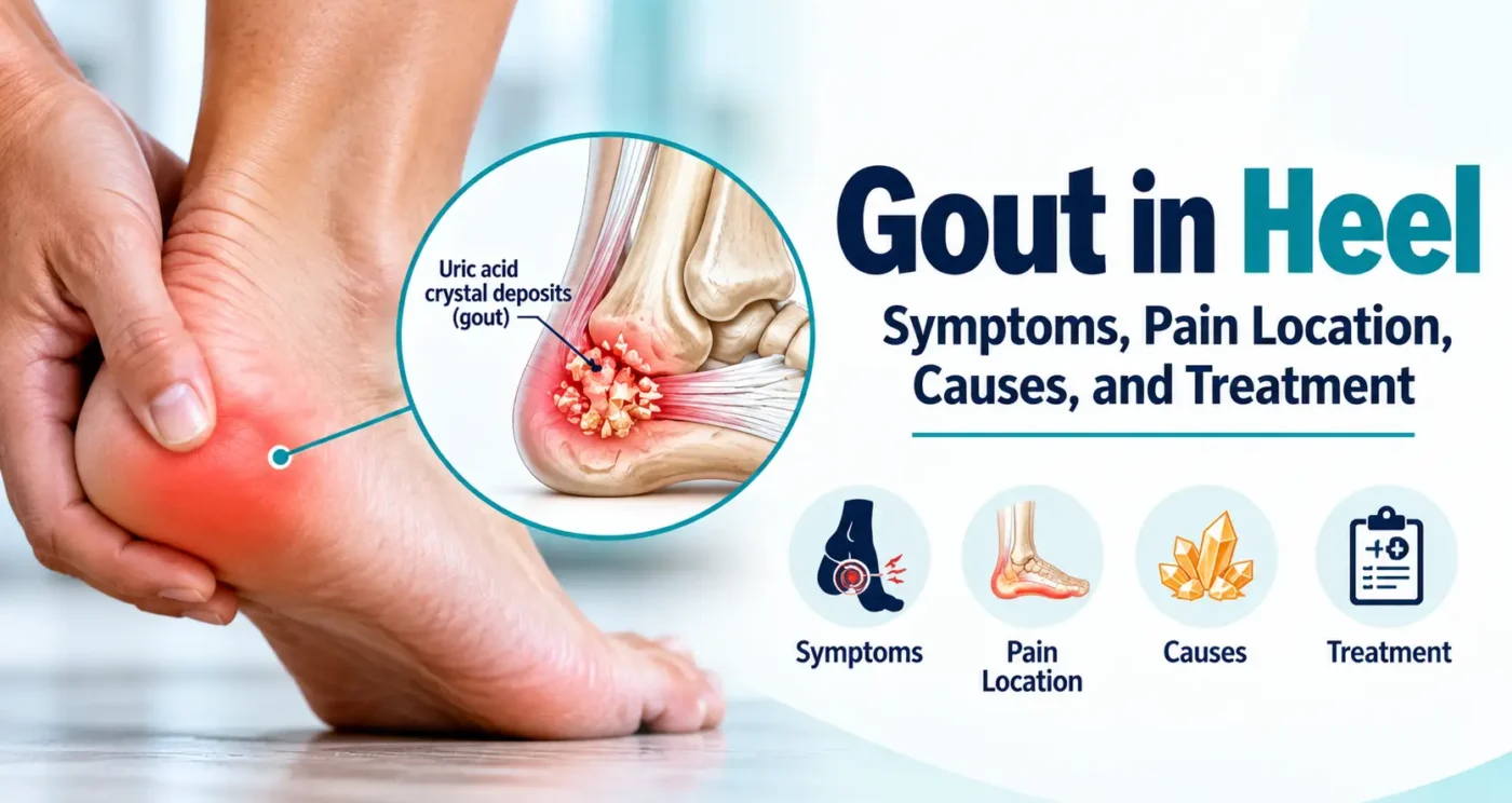
People searching for “pictures of gout in heel” or “gout in heel pictures” are often looking for a visual match. During an acute flare, photographs may show a swollen or reddish heel or ankle. In chronic tophaceous disease, images may show nodules around tendons or joints. Neither appearance is unique to gout. Some crystal deposits are deep and cannot be seen on an ordinary photograph.
A clinical photo may illustrate a possible presentation, but it cannot verify urate crystals, rule out infection, or establish the exact source of heel pain. A medically labeled illustration is often more informative than an unlabeled photo because it shows the Achilles insertion, plantar fascia, bursae and ankle structures. This article does not claim that images alone can diagnose the condition.
What Causes Gout in the Heel?
The underlying process is elevated urate over time, which can allow crystals to form in and around joints. Urate comes from normal breakdown of purines, and blood levels depend on both production and excretion, particularly through the kidneys. A high result increases gout risk but does not mean a person definitely has gout. Some people develop crystal deposits at concentrations that have been elevated for years.
Factors associated with gout include chronic kidney disease, family history, excess body weight, some diuretics, heavy alcohol intake, sugar-sweetened beverages and purine-rich foods. Certain illnesses, surgery or dehydration may precede a flare. A trigger is not always identifiable, and one food or drink rarely explains the whole condition.
The big toe is a classic first site, but longstanding hyperuricemia may be associated with deposits in less typical sites, including tendons and bursae. Heel-region symptoms should still be assessed on their own merits, even in someone with previously confirmed gout.
Gout vs. Plantar Fasciitis, Achilles Problems, and Bursitis
| Condition | Typical pattern | Useful clue |
| Gout flare | Sudden intense inflammatory pain; warmth and swelling possible | May occur at night; previous episodic attacks or tophi |
| Plantar fasciitis | Pain under heel, prominent on first steps | Often changes after walking briefly |
| Achilles tendinopathy | Pain behind heel with loading or exercise | Tender tendon insertion; may build gradually |
| Heel bursitis | Localized pain near a bursa behind heel | Shoe pressure or movement may aggravate |
| Infection | New hot, swollen, painful tissue or joint | Fever or malaise may occur; urgent assessment needed |
| Stress fracture or injury | Pain with loading, often after increased activity | Trauma or recent overuse may be relevant |
This comparison highlights patterns, not diagnostic tests. An inflamed tendon may be warm and swollen, while gout may sometimes have limited visible redness. Clinicians use examination, history and investigations when the distinction matters.
How Is Gout in the Heel Diagnosed?
Medical history and examination
A clinician will ask how quickly symptoms began, whether the pain is in the sole, tendon, joint or a lump, and whether episodes have happened before. They may review medications, kidney disease, diet, prior urate results and recent injuries. Examination can identify the point of maximum tenderness, range of motion, warmth and potential signs of infection.
Blood urate testing
A serum urate test helps assess gout risk, but a single result does not settle the diagnosis. Urate can fall during an acute flare. NICE advises repeating a low or borderline result at least two weeks after the flare resolves when suspicion remains high. Conversely, elevated urate without symptoms does not prove that heel pain is gout.
Aspiration and crystal identification
If a joint or swollen bursa contains accessible fluid, a clinician may collect a sample for microscopic examination to identify monosodium urate crystals. Fluid may also be tested for infection. This is particularly important when the diagnosis is uncertain or septic arthritis is possible. Not every heel structure is easy to aspirate, and a specialist may guide the choice of test.
Ultrasound, dual-energy CT, and other imaging
Ultrasound may reveal features suggestive of urate deposition or show tendon and bursal problems. Dual-energy CT can help identify urate deposits in selected cases. X-rays can be useful for evaluating other causes of chronic pain or damage, but they do not reliably confirm an early gout flare. Imaging should be selected for a particular clinical question.
How Is Gout in the Heel Treated?
Treatment has two goals: reduce inflammation during an active attack and, when indicated, lower urate over time so crystals dissolve and future attacks become less likely. The best medication depends on kidney function, gastrointestinal and cardiovascular risks, drug interactions, other conditions and individual preferences.
Treating an acute flare
Evidence-based first-line options include nonsteroidal anti-inflammatory drugs (NSAIDs), colchicine and corticosteroids, provided there is no contraindication. A clinician may prescribe a brief course of oral medication or select an injection in particular situations. Treatment tends to work best when started early. Do not combine anti-inflammatory drugs or change an existing prescription without medical advice.
NSAIDs may be inappropriate in some people with kidney disease, gastrointestinal bleeding risk, heart problems or certain medication combinations. Colchicine also has important interaction and kidney-related precautions; excessive dosing can be dangerous. Corticosteroids have their own risks, including changes in blood glucose. These differences matter more than the exact location of the gout attack.
Long-term urate-lowering treatment
People with frequent flares, tophi or gout-related joint damage are often candidates for urate-lowering treatment. The American College of Rheumatology recommends allopurinol as a preferred first-line option for many patients and a treat-to-target strategy, typically aiming for serum urate below 6 mg/dL. Dose adjustment and repeat blood testing are part of ongoing care. Some patients need a different target or a different medicine based on their clinical circumstances.
Starting urate-lowering therapy can temporarily increase flare risk, so anti-inflammatory prevention may be prescribed during initiation. Individuals already taking such medication should not stop it just because a flare occurs without discussing the plan with their clinician. Gout management is a long-term process, not a one-time response to heel pain.
How to Relieve Heel Pain While Awaiting Evaluation
- Rest and limit painful weight-bearing activity while the area is acutely inflamed.
- Apply a cold pack wrapped in cloth for a short period, protecting the skin and stopping if discomfort worsens.
- Use footwear that avoids direct pressure on a sore Achilles-region lump or inflamed heel.
- Drink fluids according to individual medical advice; people with fluid restrictions should follow their care plan.
- Avoid squeezing or puncturing lumps and avoid vigorous stretching of a very painful, swollen area.
- Seek a clinician’s advice about appropriate pain control rather than using leftover prescription medicines.
Ice may help symptoms but does not remove urate deposits. If swelling is accompanied by fever, significant spreading redness or an inability to bear weight, do not delay assessment to try additional home remedies.
How Long Does a Gout Flare in the Heel Last?
Untreated gout attacks often improve within one to two weeks, although the duration varies. Pain may peak early, then gradually subside. Persistent pain after visible inflammation settles does not necessarily mean crystals are still actively causing a flare; tendon injury, another heel condition or prolonged inflammation may be contributing. Recurring episodes warrant a long-term prevention discussion.
A person may feel fully normal between attacks, particularly early in the disease. That symptom-free interval is not evidence that the elevated-urate problem has been corrected. Ongoing treatment decisions rely on flare history, exam findings, blood tests and underlying health risks.
Can Gout in the Heel Make Walking Difficult?
Yes. Acute inflammation around weight-bearing joints and soft tissues can make walking painful, particularly when the heel strikes the ground or shoes rub against a swollen area. Some people change their gait to protect the foot, which can strain other areas. If walking is impossible, pain is unusually intense, or a traumatic injury is possible, assessment should not be delayed.
When the acute attack resolves, clinicians may advise a gradual return to normal activity. Persistent Achilles pain needs its own evaluation rather than repeated treatment for assumed gout.
Preventing Future Flares
Regular monitoring and an effective urate-lowering plan are often more influential than avoiding one particular food. For patients who need medication, adherence and titration to a measured urate goal help reduce future crystal accumulation. Follow-up can also address kidney health, blood pressure, cardiovascular risks and medication choices.
- Discuss recurrent flares or tophi with a primary care clinician or rheumatologist.
- Review prescription drugs, including diuretics, without stopping them independently.
- Reduce heavy alcohol intake and sugar-sweetened drinks where appropriate.
- Favor a sustainable eating pattern that includes vegetables, whole grains and suitable protein sources.
- Work toward a healthy weight gradually if recommended; crash diets can be counterproductive.
- Track flare dates, affected locations, medications used and follow-up urate results.
NIAMS also discusses the DASH eating pattern and lifestyle changes as part of gout care. These measures complement—not automatically replace—medical treatment. During an annual checkup, a medication and health-history review may help identify related risks: see MedIntelHub’s article on annual physicals (https://medintelhub.com/understanding-your-annual-physical-what-actually-gets-checked/).
When Should You See a Doctor?
Arrange medical evaluation for a first episode of unexplained heel swelling, repeated painful attacks, a new lump, ongoing pain despite rest, or difficulty walking. Gout-like symptoms arising in a new location merit reconsideration rather than automatic treatment based solely on a previous diagnosis.
Seek prompt same-day assessment for a very painful, hot, swollen joint or heel area, especially if fever, chills or a generally unwell feeling accompanies it. Septic arthritis and some soft-tissue infections can damage tissues quickly. An injury with an audible pop, loss of push-off strength or inability to bear weight also warrants urgent evaluation. Emergency care may be needed for rapidly worsening systemic illness or severe infection signs.
Frequently Asked Questions
Can you get gout in your heel?
Yes. Gout may affect joints and soft tissues around the heel, although big-toe involvement is more typical. Heel pain alone cannot confirm gout.
Can you get gout in the heel of your foot without toe pain?
Yes. A gout attack does not have to involve the big toe. The affected area and symptoms should still be evaluated for other causes.
Can gout cause heel pain when walking?
It can, because inflamed tissue may hurt when weight is placed on the foot. Achilles and plantar fascia problems can cause similar pain.
What does gout at the back of the heel feel like?
Symptoms may include tenderness, swelling, warmth or a firm deposit. Achilles tendinopathy and bursitis are important alternative causes.
Is gout on the bottom of the heel common?
Gout is not the most typical explanation for isolated plantar heel pain. Plantar fasciitis is an important possibility, especially with first-step pain.
Does gout in the heel cause redness?
Some attacks cause red or discolored skin, warmth and swelling. Redness can be subtle or absent, and infection may look similar.
Can pictures of gout in the heel confirm the diagnosis?
No. Images may show swelling or tophi but cannot identify urate crystals or rule out infection and other conditions.
Can uric acid be normal during a heel gout attack?
Yes. Blood urate may be lower during a flare. A clinician may repeat the test after symptoms settle when gout remains suspected.
Is heel gout the same as plantar fasciitis?
No. Gout is a crystal-induced inflammatory condition; plantar fasciitis affects the tissue beneath the heel. Both can cause painful steps.
How do doctors confirm gout in the heel?
They combine history and examination with selected urate testing, crystal analysis of accessible fluid, and imaging when indicated.
How long does heel gout pain last?
Many flares improve within one to two weeks, though treatment and individual circumstances can change the course.
Is ice helpful for gout heel pain?
A wrapped cold pack can help reduce discomfort during a flare. It is supportive care, not a replacement for diagnosis or appropriate medication.
Can gout deposits affect the Achilles tendon?
Yes, urate deposits can occur in tendon-related tissues, particularly in chronic tophaceous disease. Other Achilles conditions are more common causes of mechanical posterior heel pain.
Will gout in the heel return?
It can. Recurrence risk depends on urate levels, flare history and management. Long-term urate-lowering treatment may be recommended.
When is heel pain an emergency?
A hot swollen joint with fever or severe illness needs urgent assessment for infection. Severe trauma, suspected tendon rupture or rapidly worsening symptoms also require prompt care.
A Practical Way to Think About Heel Gout
A sudden, very tender, swollen heel could reflect gout—but a sore heel has several possible explanations, and no photograph or single blood test can reliably choose among them. Pain at the underside, back or sides of the heel provides clues that a clinician can combine with examination and appropriate testing. Once gout is confirmed, managing an attack and preventing future flares are different parts of care. The most useful next step is an accurate diagnosis and a treatment plan suited to the person, not the location alone.
Resources
National Institute of Arthritis and Musculoskeletal and Skin Diseases — Gout Overview
NIH overview of symptoms, causes and flares.
NIAMS — Diagnosis, Treatment, and Steps to Take
Explains clinical tests, treatment options and living with gout.
Diagnosis, Treatment, and Steps to Take
NICE — Gout: Diagnosis and Management
Guidance on urate testing, aspiration, imaging and suspected septic arthritis.
Gout: Diagnosis and Management
American College of Rheumatology — 2020 Guideline for Management of Gout
Evidence-based recommendations for flare treatment and urate-lowering therapy.
2020 Guideline for Management of Gout
American Academy of Orthopaedic Surgeons — Heel Pain
Compares plantar heel pain with posterior heel conditions.
MedlinePlus — Gout
Public-facing medical information about gout.
Editorial Guidelines & Medical Disclaimer
This article provides educational information, not an individual diagnosis or a substitute for professional medical advice. Heel pain may arise from gout, injury, infection or several other conditions, and treatment depends on examination, relevant testing and personal medical history. Do not start, stop or adjust prescription gout medicines without guidance from an appropriate healthcare professional. New hot swollen joints, fever, worsening symptoms or inability to bear weight may need urgent medical assessment. The article draws on NIH, NICE, ACR and orthopedic guidance. Recommendations can evolve; readers can review MedIntelHub’s published editorial standards below.

David Bennett
David Bennett is an author at MedIntelHub, where he creates clear, practical, and well-researched content about healthcare, medical billing, insurance, and patient education. His goal is to make complex healthcare topics easier for readers to understand and use in everyday situations. David emphasizes accuracy, transparency, and reliable sourcing, using information from trusted organizations and official healthcare resources whenever possible. Content published by David is intended for educational purposes and should not replace advice from qualified medical, legal, or healthcare professionals.