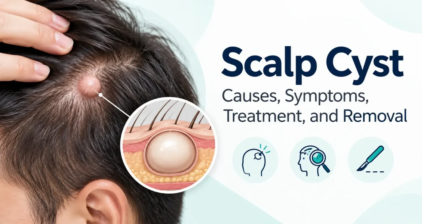
A small bump on the scalp can go unnoticed until a comb catches it or a fingertip finds it while washing hair. Many of these lumps turn out to be harmless cysts. Others are not cysts at all, and the difference is not always clear from appearance. A scalp cyst is often painless and slow-growing, but changes in size, tenderness, or the overlying skin deserve attention.
Pilar cysts, which arise from hair follicles, are especially associated with the scalp. Epidermoid cysts can also occur there. Neither label can be confirmed simply by looking in a mirror, and treatment depends on the actual lesion, its symptoms, and whether its diagnosis is certain. This guide explains what a cyst on the scalp may feel like, how clinicians distinguish different lumps, and what removal and recovery usually involve.
What Is a Scalp Cyst?
A cyst is a sac-like structure beneath or within the skin that contains material produced by its lining. Scalp cysts commonly hold keratin, the structural protein found in skin, hair, and nails. They are not simply pockets of grease or ordinary liquid. A cyst often grows gradually as material accumulates inside its wall.
The word “cyst” describes a type of growth, not an explanation of every lump on the head. A clinician may examine the lesion to decide whether it is a pilar cyst, an epidermoid cyst, an inflamed follicle, a lipoma, or something requiring a different workup. Cleveland Clinic pilar cyst describes the usual scalp presentation of pilar cysts and their follicular origin.
What Does a Cyst on the Scalp Look and Feel Like?
A typical uncomplicated pilar cyst is a smooth, rounded lump beneath intact skin. It may feel firm, shift slightly beneath the fingertips, and have no obvious opening on its surface. Hair often grows over it normally. One person may have a single small nodule; another may have several. The lump may be noticeable only when lying on a pillow, getting a haircut, or wearing a helmet.
Epidermoid cysts may feel similar, but some have a small central pore or punctum. A cyst can become red, swollen, or tender if its contents leak into surrounding tissue or if bacterial infection develops. Skin tone affects the appearance of redness: inflammation can look pink or red on lighter skin and darker, purplish, or subtly discolored on deeper skin.
Pain, rapid enlargement, bleeding, persistent drainage, or a growth that feels fixed to underlying structures should not be dismissed as normal cyst behavior. Those findings call for a clinical examination rather than home removal.
Types of Scalp Cysts
Pilar (Trichilemmal) Cysts
Pilar cysts develop from the outer root sheath of a hair follicle. They are strongly associated with the scalp, where hair follicles are plentiful. They typically contain compact keratin and lack the central opening sometimes seen in epidermoid cysts. Many are asymptomatic for years, and a tendency to develop multiple pilar cysts can run in families.
Pilar cysts are generally benign. Rarely, a lesion resembling a long-standing pilar cyst may be a proliferating trichilemmal tumor, particularly when it enlarges unusually quickly or develops ulceration. This possibility does not mean a stable scalp cyst is likely to be cancer, but it is one reason clinicians may recommend examination and pathology for atypical growths. DermNet pilar cyst; DermNet proliferating cyst.
Epidermoid Cysts
Epidermoid cysts arise when epidermal cells accumulate within a sac under the skin. They can appear on many body sites, including the scalp. A small punctum may be visible; the contents may be thick and have an unpleasant smell if released. Cysts can become inflamed after rupture even without bacterial infection. Complete removal of the sac reduces the chance that the same cyst returns. DermNet epidermoid cyst.
Other Cyst-Like Lumps
A dermoid cyst, especially in a younger patient, may arise from developmental tissue and sometimes needs imaging depending on its location. Scalp lumps may also represent lipomas, abscesses, inflamed hair follicles, or other skin tumors. A lump close to a bony suture, present since infancy, or apparently fixed in place deserves individualized assessment.
Pilar vs. Epidermoid Cysts: At a Glance
| Feature | Pilar cyst | Epidermoid cyst |
| Common location | Especially scalp | Face, trunk, scalp, and other sites |
| Origin | Outer root sheath of hair follicle | Epidermal cells in a cyst lining |
| Typical surface | Usually no central punctum | May have visible central punctum |
| Contents | Dense keratin | Keratinous material |
| Usual course | Slow-growing and often painless | Slow-growing and often painless |
| If troublesome | Observation or complete removal | Observation or complete removal |
These patterns are useful clues, not a substitute for examination or tissue diagnosis when indicated. Mayo Clinic cyst symptoms; DermNet epidermoid cyst.
What Causes a Scalp Cyst?
Pilar cysts are associated with the way cells in the hair-follicle sheath grow and produce keratin. In some families, the tendency to form them is inherited. Many people cannot identify a triggering event. Having a pilar cyst does not mean the scalp is dirty or that hair was washed incorrectly.
Epidermoid cysts form when epidermal cells become trapped under the surface and continue producing keratin. Sometimes this follows disruption of a hair follicle or skin injury, although a definite trigger often is not apparent. Cysts are not generally caused by touching the scalp too frequently, and ordinary shampoo choices are unlikely to explain them.
A person may discover a cyst after a haircut or after pressure from headwear makes it uncomfortable. The timing of discovery is not necessarily the timing of formation.
What Symptoms Can a Scalp Cyst Cause?
Most uncomplicated scalp cysts produce little besides a palpable lump. They may create pressure when resting the head, brushing hair, or using headphones. A larger lesion can stretch nearby skin and become more noticeable.
Inflammation can change the picture: swelling, tenderness, warmth, and surface discoloration may develop over a short period. A ruptured cyst can drain thick keratinous material. Symptoms should be judged in context, because a painful lump can also be an abscess or a different condition.
Can a Scalp Cyst Become Infected?
Yes, a cyst can develop a bacterial infection, but infection and inflammation are not synonyms. If the cyst wall ruptures, keratin released into surrounding tissue can provoke a pronounced inflammatory response. The area may become painful and red even when bacteria are not the main cause. This distinction matters because antibiotics do not remove the sac and are not automatically helpful for every inflamed cyst. DermNet pilar cyst.
Signs suggesting infection include increasing warmth, spreading redness or discoloration, pus, worsening pain, and sometimes fever. A clinician may need to examine the area, drain a collection if present, or prescribe medication when bacterial infection is suspected. Do not try to open an inflamed lump with a needle at home.
Is a Scalp Cyst Dangerous or Cancerous?
Most typical pilar and epidermoid cysts are benign. The greater everyday risks are irritation, inflammation, rupture, and recurrence. However, a new scalp lump should not automatically be labeled a cyst without assessment. Some skin cancers and unusual follicular tumors can resemble benign nodules, especially early on.
A lesion that enlarges rapidly, bleeds without clear injury, ulcerates, becomes firmly attached, or repeatedly changes after treatment deserves prompt evaluation. A dermatologist or surgeon may recommend removal and microscopic examination to confirm the diagnosis. DermNet scalp lesions provides a wider differential diagnosis for scalp growths.
How Is a Scalp Cyst Diagnosed?
Clinicians usually begin with a history and examination. They may ask how long the bump has been present, whether it has grown or drained, whether similar cysts occur in the family, and whether it causes pain. The examination considers size, skin changes, mobility, consistency, and whether there are other lesions.
For a straightforward superficial cyst, imaging is often unnecessary. Ultrasound can be useful for an uncertain lump; CT or MRI may be considered for unusually deep, fixed, large, recurrent, or anatomically concerning lesions. These tests are selected for the individual situation rather than ordered routinely for every scalp bump.
If a cyst is removed, the tissue may be sent for histopathology. A pathology report can confirm its type and help rule out a different growth, especially when the appearance was atypical.
Scalp Cyst Treatment Options
Observation
A small, stable, painless cyst often needs no immediate treatment after a clinician has assessed it. Some people choose to leave a lesion alone if it causes no functional or cosmetic concern. Follow-up becomes more important if it changes noticeably, causes pressure, or begins to drain.
Managing Inflammation or Infection
A clinician may manage an inflamed cyst differently from an uncomplicated one. An active infection or intense swelling can make definitive excision harder; treatment might first address the inflammation or any abscess. Antibiotics are reserved for appropriate circumstances, not prescribed simply because a cyst is present. For selected epidermoid cysts, an injection to reduce inflammation may be discussed. Mayo Clinic cyst treatment.
Surgical Removal
For a symptomatic or recurring cyst, planned surgical excision is often definitive. The goal is removal of the cyst and its lining, not just emptying its contents. If the wall is left behind, material can accumulate again. The method and timing depend on lesion size, location, inflammation, and the clinician’s assessment.
When Should a Scalp Cyst Be Removed?
Removal may be worth discussing when the cyst repeatedly catches on a comb, hurts when pressure is applied, grows, becomes inflamed more than once, or causes substantial distress. Diagnostic uncertainty is another reason to consider biopsy or excision. Conversely, a small, uncomplicated, confirmed cyst does not require surgery solely because it exists.
The decision also involves scarring, wound healing, local anesthesia, possible hair trimming, and recurrence risk. A specialist can explain whether monitoring, office excision, or another approach is most appropriate.
How Is Scalp Cyst Removal Performed?
Many superficial scalp cysts can be removed in an outpatient clinic under local anesthetic. The clinician cleans the skin, numbs the area, makes an appropriately sized incision, and carefully separates the cyst from surrounding tissue. They try to remove the lining intact when possible. The wound may be closed with stitches and covered with a dressing.
Some cysts can be removed with a relatively small opening, but technique depends on the lesion. If the cyst is very inflamed, ruptured, or adherent, removal may be more difficult. Larger or unusual lesions can require specialist management. The extracted tissue may be examined in a laboratory.
Whether a patch of hair needs trimming is case-dependent. Not everyone needs the surrounding scalp shaved. Discuss this beforehand if appearance or work obligations matter to you.
Recovery After Scalp Cyst Removal
The scalp has a rich blood supply, and minor oozing early after surgery may occur. Your clinician will give specific instructions about dressings, when to wash your hair, which activities to avoid, and whether stitches need removal. Instructions vary with wound size, location, and closure method, so a generic timeline should not override the treating team’s directions.
Tenderness and mild swelling can occur in the first days. A small scar is possible, and hair may be temporarily disturbed near the incision. Seek advice if pain increases rather than improves, the wound opens, bleeding is difficult to control, or drainage, fever, or spreading redness develops.
Aftercare can be simpler for a small excision than for a larger or previously infected lesion. Ask whether the tissue will undergo pathology and how the result will be communicated.
Can a Scalp Cyst Come Back After Removal?
Recurrence is possible, particularly if part of the cyst wall remains. That is why simply squeezing out contents or draining a cyst does not provide the same long-term result as complete excision. New cysts can also form in other scalp follicles, especially in someone prone to pilar cysts. These are new lesions rather than necessarily a return of the original one.
A recurring bump at a surgical scar should be checked, particularly if it is enlarging or behaving differently from the earlier lesion. DermNet epidermoid cyst describes the role of intact capsule removal in limiting recurrence.
Can You Treat a Cyst on the Scalp at Home?
Home care cannot reliably dissolve a true pilar or epidermoid cyst. If a previously examined lesion is mildly uncomfortable, avoiding pressure from combs or helmets may help prevent further irritation. Gentle scalp hygiene is reasonable. Do not puncture, squeeze, or attempt to cut out the cyst; these actions can injure the skin, push material into tissue, and cause infection or scarring.
Warm compresses may soothe some tender superficial lumps, but they do not remove a cyst wall and should not delay assessment of worsening redness, pain, or fever. Do not apply harsh chemical products or unproven “drawing salves” to an open or inflamed area.
Scalp Cyst vs. Other Scalp Lumps
| Type of lump | Possible clue | Why assessment matters |
| Pilar cyst | Smooth, rounded, often scalp-based | May be monitored or excised |
| Epidermoid cyst | May have a central punctum | Can inflame or rupture |
| Lipoma | Often softer, rubbery, movable | Different tissue origin and management |
| Abscess | Painful, warm, swollen, may contain pus | May need prompt drainage and infection care |
| Inflamed follicle | Small tender bump centered on a hair | Usually managed differently from a cyst |
| Other skin tumor | Changing, bleeding, ulcerated or atypical lesion | May need biopsy or specialist evaluation |
No single sign on this table confirms a diagnosis. Several conditions overlap, and professional assessment matters when the lump is new, atypical, or changing.
When Should You See a Doctor?
Arrange a routine appointment for a new scalp lump even when it is painless, particularly if you do not know what it is. A visit is also reasonable if a known cyst is growing, affecting daily activities, or repeatedly inflamed. During a Annual physical examination, bring up any new or changing skin growth directly rather than assuming it will be examined automatically.
Seek prompt care for increasing pain, warmth, pus, spreading inflammation, or fever. A rapidly growing, firm, ulcerated, or bleeding lesion should be evaluated rather than treated as a harmless cyst. Severe illness, extensive rapidly spreading infection, or concerning systemic symptoms require urgent medical assessment.
Frequently Asked Questions
What is a scalp cyst filled with?
Most pilar and epidermoid cysts contain keratin, a protein associated with skin and hair, rather than ordinary oil or clear fluid.
Is a cyst on the scalp common?
Pilar cysts are especially associated with the scalp. A lump may be a different condition, so an examination helps identify it.
Are scalp cysts usually painful?
Many are painless. Pain can occur with pressure, rupture, inflammation, or infection.
Can a scalp cyst disappear on its own?
A true cyst often persists because its lining remains. Changes in swelling do not necessarily mean the cyst has disappeared.
What causes multiple scalp cysts?
Some people inherit a tendency to develop pilar cysts, although multiple bumps still merit examination.
Can a scalp cyst cause headaches?
Pressure or tenderness from a large or inflamed scalp lesion may be uncomfortable, but a headache should not automatically be attributed to a cyst.
Can a scalp cyst become cancerous?
Most are benign. Unusual, rapidly enlarging, ulcerated, or bleeding lesions need assessment because other tumors can resemble cysts.
Does a scalp cyst need surgery?
Not always. Observation may be reasonable for a confirmed, stable, asymptomatic cyst; removal is considered for symptoms, recurrence, or diagnostic uncertainty.
Does scalp cyst removal hurt?
Local anesthetic usually numbs the procedure area, although injection and later soreness can cause temporary discomfort.
Will the doctor shave my hair for cyst removal?
Sometimes a small area is trimmed, but the need depends on the cyst and surgical method.
How long does healing take?
Healing varies with wound size, closure, and medical factors. Follow the treating clinician’s instructions about dressings, washing, and stitches.
Can a cyst grow back after drainage?
Yes. Draining contents without removing the cyst lining can allow the lump to refill.
Should I pop a scalp cyst?
No. Popping or puncturing it can increase inflammation, infection risk, and scarring.
Is a red scalp cyst always infected?
No. Ruptured keratin can cause inflammation without bacterial infection; a clinician can evaluate whether antibiotics or drainage are appropriate.
Which doctor removes scalp cysts?
Primary-care clinicians, dermatologists, and surgeons may evaluate or remove scalp cysts, depending on size, location, complexity, and local services.
What to Remember About a Scalp Cyst
A slow-growing, painless scalp lump is often benign, and pilar cysts are a familiar explanation. But “cyst” should be a diagnosis rather than a guess. Changes in size, pain, drainage, or skin appearance call for an assessment, while sudden signs of infection deserve prompt attention. If removal is appropriate, ask whether the complete cyst wall will be removed, how the wound should be cared for, and whether pathology is planned.
Resources
DermNet — Trichilemmal (Pilar) Cyst
Description of follicular origin, characteristic pathology, inflammation, and treatment. DermNet pilar cyst
Cleveland Clinic — Pilar Cyst
Patient-oriented explanation of scalp presentation, removal, recurrence, and warning signs. Cleveland Clinic pilar cyst
DermNet — Epidermoid Cyst
Clinical differences, complications, and the role of complete cyst excision. DermNet epidermoid cyst
Mayo Clinic — Epidermoid Cyst Diagnosis and Treatment
Clinical approach to observation, inflammation, drainage, and surgical removal. Mayo Clinic cyst treatment
DermNet — Scalp Tumors and Cysts
Overview of other conditions that can present as scalp nodules. DermNet scalp lesions
DermNet — Proliferating Trichilemmal Cyst
Describes a much less common growth requiring diagnostic assessment and excision. DermNet proliferating cyst
NHS — Skin Cyst
General public guidance on skin cysts and when to seek clinical evaluation. NHS skin cyst
Editorial Guidelines & Medical Disclaimer
This MedIntelHub article provides general educational information and does not diagnose an individual scalp lump or replace evaluation by a qualified healthcare professional. A new, changing, painful, or bleeding lesion may require clinical examination and, sometimes, testing or tissue pathology. Treatment decisions, including medicines and surgical procedures, depend on personal circumstances; do not attempt to puncture or remove a cyst yourself or change prescribed treatment without professional guidance. Seek prompt care for concerning infection symptoms and urgent care for rapidly progressing severe illness. This article draws on established dermatology and clinical resources; recommendations can change. Read the MedIntelHub editorial policy for our research and review approach. When applicable, insurance and billing policies also vary.

David Bennett
David Bennett is an author at MedIntelHub, where he creates clear, practical, and well-researched content about healthcare, medical billing, insurance, and patient education. His goal is to make complex healthcare topics easier for readers to understand and use in everyday situations. David emphasizes accuracy, transparency, and reliable sourcing, using information from trusted organizations and official healthcare resources whenever possible. Content published by David is intended for educational purposes and should not replace advice from qualified medical, legal, or healthcare professionals.